

Jon M. Chang
Popular Mechanics
January 29, 2013
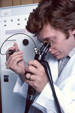
You can get an X-ray to see your bones, an MRI to see your brain, and a CT scan to see virtually everything else. But getting a glimpse of the gastrointestinal tract (the esophagus, stomach, and intestines) is a little more invasive. Endoscopes have been able to provide doctors with a picture of their patients’ insides, but they have their limitations: For instance, the person performing the endoscopy needs extensive training on guiding the camera down the patient’s throat. It’s also a time-intensive procedure that requires the patient to be sedated.
A group of doctors and engineers working at both Ninepoint Medical and Massachusetts General Hospital developed a new endoscope that gets around these hurdles by thinking small. As reported in a recent study in Nature Medicine, they miniaturized the endoscope to fit the imaging equipment into a clear capsule patients can swallow. One end of the capsule is attached by a 1-mm-wide cable tethered to a console near the patient. When it reaches the stomach, the doctor can reel it back in and take snapshots of the GI tract on its return trip.
Michalina Gora, lead author of the study and a research fellow at Massachusetts General Hospital, says that one of her biggest concerns was how patients coped with a long string dangling in their throat. “Sometimes, the procedure triggered a gag reflex in the patient,” she says. “This can be taken care of just by breathing steadily, in and out. It opens up the GI tract’s sphincters and allows the endoscope to pass through.” Overall, patients prefer the swallowable endoscope to the standard version because the scan takes less time (about 5 minutes) and doesn’t require sedation.
The Emergency Election Sale is now live! Get 30% to 60% off our most popular products today!